 My Cart



 My Cart





Product Overview

Product List

Product Classification

Quality System

Case Data
Stable Overexpression Cell Line Products
Product Overview
Overexpression cell lines refer to cell lines where target genes are efficiently integrated into the host cell’s chromosomes using technologies such as lentiviruses and electroporation, enabling long-term, stable, and high-level expression of the target gene. Based on a high-throughput cell line development platform, Sanyou Bio's stable overexpression cell lines are based on an AI-assisted codon optimization system and its proprietary high-expression vector system, which can increase the expression of the target protein by hundreds to several hundred thousand times, effectively enhancing experimental signals and improving detection sensitivity.
With the established high-throughput cell line construction platform, Sanyou Bio has over 5,000+ cell line construction experiences. Currently, we have stocked more than 800 types of stable overexpression cell line products, covering categories such as overexpression cell lines, reporter gene cell lines, and EGFP overexpression cell lines. All products comply with internal release standards.
Stable overexpression cell lines are suitable for gene function research, drug screening, signaling pathway research, and various mechanism validation scenarios. They can serve as reliable and high-performance cell models to accelerate basic research and drug development processes.
Product List
Sanyou Bio's cell line products include eukaryotic overexpression cell lines, reporter gene cell lines, and EGFP overexpression cell lines. All cell line products are in stock to ensure fast delivery. Every cell line has undergone strict stability verification, flow cytometry validation, and possesses clear and traceable source information, fully meeting the requirements of scientific research projects and application compliance.

Eukaryotic Overexpression Cell Lines

EGFP Overexpression Cell Lines

Reporter Gene Cell Lines

Tumor Cell Lines Validated by Modeling
Product Classification
Showing 1 to 20 article,A total of 20 Bar result
- 1
Total Pages: 0
Quality System
Every cell line undergoes multiple quality inspections to ensure compliance with the highest quality standards. Our production process adheres to the ISO 9001 quality management system, with strict full-process monitoring implemented from raw material procurement to finished product delivery.
Stability Test: ≥16 PDL
Mycoplasma Detection: Negative
Flow Cytometry Detection Positive Rate: ≥70%
Quality Test Report
Each product is accompanied by a detailed quality test report, including flow cytometry analysis, mycoplasma detection, etc.
View Sample Report >
Case Data
PD-1 / PD-L1 Luciferase Reporter Gene Cell Line
Eukaryotic Jurkat Overexpression Cell Lines
Tumor Cell Line Validated by Modeling
PD-1 / PD-L1 Luciferase Reporter Gene Cell Line
1. Product Description
The engineered Human PD-1 (Luc) Jurkat Reporter Cell stably expresses an NFAT response element-driven luciferase system and full-length human PD-1, which can be used to evaluate the activity of PD-1/PD-L1 blockers.
When co-cultured with target cells expressing human PD-L1 / anti-CD3 scFv, the interaction between PD-1 and PD-L1 inhibits the TCR signaling pathway and NFAT-mediated luciferase activity. In contrast, anti-PD-1/PD-L1 antibodies block this interaction to relieve the inhibition, activate the TCR pathway, and induce luciferase expression.
When co-cultured with target cells expressing human PD-L1 / anti-CD3 scFv, the interaction between PD-1 and PD-L1 inhibits the TCR signaling pathway and NFAT-mediated luciferase activity. In contrast, anti-PD-1/PD-L1 antibodies block this interaction to relieve the inhibition, activate the TCR pathway, and induce luciferase expression.
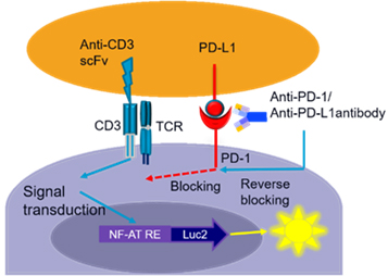
Schematic Diagram of PD-1 / PD-L1 Luciferase Reporter Gene Detection System Principle
2. Validation of Atezolizumab Antibody (Anti-PD-L1) Blocking Activity
Purpose:To verify the blocking effect of Atezolizumab on the downstream signaling pathway of the PD-1/PD-L1 luciferase reporter gene cell line.
Method:Serially diluted Atezolizumab was co-incubated with aAPC cells and human PD-1 (Luc) Jurkat Reporter Cells for 6 hours. After adding the chromogenic substrate, the optical density (OD) at 492 nm was detected.
Result: Atezolizumab showed a significant blocking effect with an IC₅₅ of 0.55 nM。
Conclusion: Atezolizumab has blocking activity, and the cell line was successfully constructed.
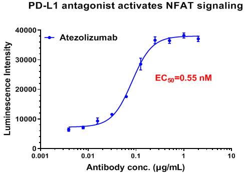
Atezolizumab Antibody (Anti-PD-L1) Activates TCR Signaling


